Surgeries and Procedures: Retinopathy of Prematurity
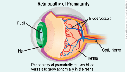
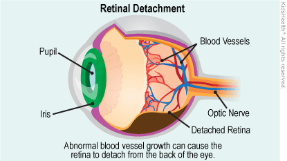
Retinopathy of prematurity (ROP) is a disease of premature babies that causes abnormal blood vessels to grow in the retina, the layer of nerve tissue in the eye that enables us to see. This growth can cause the retina to detach from the back of the eye, leading to blindness.
Some cases of ROP are mild and correct themselves, but others require surgery to prevent vision loss or blindness. Surgery involves using a laser or other means to stop the growth of the abnormal blood vessels, making sure they don’t pull on the retina.
Because there are varying degrees of ROP, the surgical approach used can differ for each case. The more you know about retinopathy of prematurity and your baby’s surgery, the easier the experience is likely to be for you.
About ROP
Located in the inner membrane of the eye, the retina is a layer of nerve tissue that captures light from an object and converts it to electrical impulses, then sends these impulses to the brain. For the retina to work properly, it must receive blood from a network of blood vessels.

When blood vessels grow abnormally and randomly, they become fragile and tend to leak or bleed. This causes scarring of the retina. When the scars shrink, they pull on the retina, causing it to detach from the back of the eye. Since the retina is a vital part of vision, detachment of the retina will cause blindness.
ROP can stop or reverse itself at any point, so it often resolves as a baby grows. But if the disease has progressed to the point of scarring, the retina will begin to detach.
ROP has no signs or symptoms. The only way to detect it is through an eye examination by an ophthalmologist.

Causes
Normally, blood vessels grow from the center of a developing baby’s retina 16 weeks into the mother’s pregnancy, and then branch outward and reach the edges of the retina 8 months into the pregnancy. In babies born prematurely, normal retinal vessel growth may be disrupted and abnormal vessels can develop, which can cause leaking and bleeding in the eye.
About ROP Surgery
The goal of ROP surgery is to stop the growth of abnormal blood vessels by focusing treatment on the peripheral retina (the sides of the retina) to preserve the central retina (the most important part of the retina). ROP surgery involves scarring areas on the peripheral retina to stop the abnormal growth and eliminate pulling on the retina.
Since surgery focuses treatment on the peripheral retina, these areas will be scarred and some amount of peripheral vision may be lost. However, by preserving the central retina, the eye will still be able to perform vital functions like seeing straight ahead, distinguishing colors, reading, etc.
The most frequently used methods of ROP surgery are:
- laser surgery, the most common type of ROP surgery, in which small laser beams are used to scar the peripheral retina (also called laser therapy or photocoagulation)
- cryotherapy, where freezing temperatures are used to scar the peripheral retina. For many years, cryotherapy (also called cryosurgery) was the accepted method of ROP surgery, but it has been all but replaced by laser therapy.
For more advanced cases of ROP where retinal detachment has occurred, these methods are used:
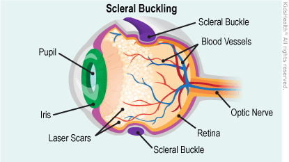
- scleral buckling, which involves placing a flexible band, usually made of silicone, around the circumference of the eye. The band is placed around the sclera, or the white of the eye, causing it to push in, or “buckle.” This, in turn, causes the torn retina to push closer to and remain against the outer wall of the eye.
- vitrectomy, a complex surgery that involves replacing the vitreous, or the clear gel in the center of the eye, with a saline (salt) solution. Removing the vitreous allows for the removal of scar tissue and eases pull on the retina. This stops the retina from pulling away.
Your baby’s ophthalmologist will determine and discuss with you which ROP surgery method is best.
Preparing for Surgery
Once your child is admitted to the hospital, you’ll have to fill out some paperwork and provide basic information, including:
- your baby’s gestational age at birth
- your baby’s birth weight
- name and phone number of your pediatrician
- your insurance provider
You’ll wait for surgery in the pre-operative area within the hospital, and your baby will be given an ID bracelet. A nurse will check some of your baby’s vital signs and put in place these monitors:
- a blood pressure monitor, which periodically checks your baby’s blood pressure throughout the procedure. It is simply a cuff that fits around the arm.
- a pulse oximeter, which measures the level of oxygen in the blood and monitors heart rate. A pulse oximeter resembles a bandage and is placed on a baby’s fingertip.
- a heart monitor, which checks the rhythm and rate of the heartbeat. A heart rate monitor is attached to a series of small metal tabs (called electrodes) that are connected to a machine that records the activity of the heart. The electrodes have adhesive backs that stick to the skin and are placed on the chest.
Depending on the availability of a surgical room, you and your baby might have to wait for a little while for the surgery to begin.
Talking to the Pediatric Ophthalmologist
Your baby’s pediatric ophthalmologist will probably describe the procedure and answer any questions you might have. This is a good time to ask questions about anything regarding the procedure that you don’t understand.
Once you feel comfortable with the information and your questions have been fully answered, you’ll be asked to sign an informed consent form, stating that you understand the procedure and its risks and give your permission for the surgery.
Anesthesia
ROP surgery is typically performed either under general anesthesia (medication that keeps the patient in a deep sleep-like state) or deep sedation (medication that makes the patient unaware of the procedure but not as deeply sedated as general anesthesia).
Laser surgery and cryotherapy are performed under deep sedation, while scleral buckling and vitrectomy are performed under general anesthesia. It’s very important to follow your doctor’s instructions on when to have your child stop eating or drinking (for babies on breast milk only, this is typically 2 hours before the procedure; for toddlers and older children, it may be up to 8 hours before).
The anesthesiologist or certified registered nurse anesthetist (CRNA) will explain the details about the type of anesthesia to be used. Feel free to ask the anesthesiologist or CRNA any questions you might have. Once you feel comfortable about the procedure, you’ll be asked to sign another informed consent form, this time authorizing the use of anesthesia.
During the Surgery
Although there is no cutting or stitching involved in laser surgery or cryotherapy, all surgical procedures for retinopathy of prematurity require that the baby be given sedation and pain medication or general anesthesia.
Laser surgery and cryotherapy are usually done at the child’s bedside with sedation and pain medication. Since scleral buckle and vitrectomy surgeries require general anesthesia, they are performed in an operating room. For all procedures, your baby’s breathing and heart rate will be closely watched during the surgery.
Your child will receive eye drops to dilate the pupil(s) before each procedure. During the surgery, a tool called an eyelid speculum will be gently inserted under the eyelids to keep them from closing.
Whether a child will be kept in the hospital is determined by his or her age at the time of surgery in addition to medical condition. In most hospitals, any children undergoing surgery before 52 weeks of gestational age will be admitted for 23 hours’ observation after the procedure.
The eye will be covered with a patch after scleral buckling and vitrectomy, but not after laser or cryotherapy.
Laser Surgery
The purpose of both laser surgery and cryotherapy is to create scar tissue on the peripheral retina. The scarred peripheral retina will no longer stimulate growth of abnormal vessels. As a result, normal retinal vessels will extend into the area where they were absent before the treatment. Although the scarred area of the peripheral retina will no longer work, the central retina (the most important part of the retina) will work fine and vision will be preserved.
Laser surgery is the most common type of ROP surgery and uses a beam of light to create scar tissue on the peripheral retina. Using the laser, the ophthalmologist aims beams of light at the peripheral retina and burns, then scars, the area where abnormal blood vessels have not yet reached.
Laser surgery usually takes about 30-45 minutes for each eye. After the surgery, you’ll probably notice that your baby’s eyes and eyelids are a little red. Also, you might see some swelling around the eyelids. This is normal and can last from a few days to a few weeks. The eye will not be covered and your baby will receive eye drops for a week.
Cryotherapy
Cryotherapy freezes and scars the peripheral retina to stop abnormal blood vessels from growing.
For a cryotherapy procedure, a metal probe that has been exposed to liquid nitrogen (an extremely cold gas) is placed on the sclera, or white of the eye. The freezing temperature then quickly travels to the peripheral retina and freezes and scars the area.
Cryotherapy treatment takes about 30-45 minutes. You will notice redness in your baby’s eye and possibly around the eyelid, as well as some swelling. The eye will not be covered and your baby will receive eye drops.
Scleral Buckling
Scleral buckling is a procedure reserved for ROP cases in which the retina has started to detach. The concept involves wrapping a band or “buckle” around the side of the eye (in other words, not around the lens). This band forces the retina to remain in place. This “buckling” reduces the pressure and pulling on the retina. This procedure is performed by a retinal surgeon.
The surgeon first uses laser surgery or cryotherapy to destroy the abnormal blood vessels. After that, a band (commonly made of silicone) is wrapped around the sclera and over the detaching retina. If there is too much fluid under the detached retina, the surgeon might make a small incision to drain it. Then, the band is stitched in place.

Scleral buckling can take 1-2 hours and your baby will have redness in the eyes and around the eyelids, as well as some swelling in that area. The eye will be covered with a patch until it heals.
Patients who undergo scleral buckling should have the buckle examined every 6 months, or more frequently if there is a problem. The buckle will be removed if there is a sign that it is too tight or causing a problem. Eventually, it will be permanently removed (months or years later) to accommodate the growing eye.
Vitrectomy
Vitrectomy involves removing the eye’s vitreous (the clear gel in the center of the eye) and replacing it with a saline solution that functions much like the real thing. Vitrectomy allows a retinal surgeon to remove scar tissue from the inside of the eye and relieve pulling on the retina.
Using a microscope and special lenses placed on the eyeball to get a better view of the back of the eye, the surgeon will use a scalpel to make several small incisions on the sclera. Tiny surgical tools are inserted through the incisions, including a fiber-optic tool for light and laser treatment, a small cutting tool, and an infusion line (a tube that maintains pressure so the eye doesn’t lose its shape).
The cutting tool is used to remove the vitreous and eliminate any scar tissue, blood, or debris in the eye. Then, using the infusion line, the vitreous is replaced with a saline solution.
All incisions are then closed with sutures (stitches) that will dissolve on their own. A vitrectomy can take several hours. Your baby will have redness in the eyes and around the eyelids, as well as some swelling in that area. The eye will be covered with a patch until it heals.
Recovery time can take several weeks, and a baby who undergoes vitrectomy will need frequent eye exams.
After the Procedure
If admission to the hospital isn’t necessary, you’ll be able to take your child home about an hour after the procedure. Follow-up care for ROP surgery includes giving your child eye drops (to prevent infection) for at least a week.
To make sure the eyes are healing properly and that ROP hasn’t returned, eye exams should be scheduled based on instructions from the ophthalmologist. This is usually every 1-2 weeks. For scleral buckling, the ophthalmologist must examine the buckle every 6 months to account for your child’s growing eye.
Benefits
Treating retinopathy of prematurity helps preserve a child’s vision. If the disease isn’t addressed, abnormal blood vessels can cause the retina to detach, permanently damaging the eyes and causing blindness.
Risks and Complications
The goal of surgery for retinopathy of prematurity is to stop the progression of the disease and prevent blindness. Although ROP surgery has a good success rate, not all babies respond to treatment. Up to 25% of babies who have ROP surgery might still lose some or all vision.
In some cases, laser surgery or cryotherapy isn’t successful (meaning the abnormal blood vessels continue growing) and might have to be repeated. If the growth continues, scleral buckling or vitrectomy might have to be attempted. However, it’s important to know that surgery for advanced stages of ROP doesn’t guarantee that the retina will re-attach and sight will be restored.
Keep in mind that for all types of ROP surgery, a degree of your child’s peripheral (side) vision will be lost. And even if the ROP has stopped progressing, vision can still be affected in the following ways:
- strabismus: the eyes aren’t aligned correctly (one example is a “crossed eye”)
- amblyopia: one eye is weaker than the other (sometimes called “lazy eye”)
- myopia: nearsightedness or difficulty seeing distant objects
- glaucoma: increased pressure on the eye that can damage the optic nerve and cause vision loss or blindness
- retinal detachment: the retina detaches from the eye, causing vision loss or blindness
Since some vision loss and complications can occur, any child that has had ROP surgery should have regular, yearly eye exams well into adulthood.
