Congenital Pigmented Moles (Congenital Nevi)

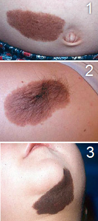
Fig. 1. Small congenital nevus. This is a benign-appearing, small congenital mole with uniform pigmentation & borders.
Fig. 2. Intermediate-size congenital nevus. This lesion has regular borders & pigmentation with long, luxuriant hair growth within the mole.
Fig. 3. A benign-appearing mole
Congenital nevus refers to a brown birthmark which is a common skin growth composed of special pigment-producing cells called nevomelanocytes. These cells are related to pigment producing cells normally found in the skin. The size of the nevus may vary from a small one-inch mark to a giant birthmark covering half of the body or more. The size and location of the nevus will determine the risk of the development of melanoma, and ease of surgical removal.
The distinction between congenital nevi and other forms of acquired moles is usually made on clinical examination. Any nevus larger than 1.5 cm is likely to be congenital. Some distinguishing pathological features, from skin biopsy, can help diagnose congenital nevi if necessary.
How common are congenital moles?
Small congenital pigmented moles (brown birthmarks) are present in 1 percent of all newborn babies. Giant congenital moles (larger than 8 inches) are rare, found in fewer than one in 20,000 newborn infants.
Why are they special?
The risk of change to melanoma, a dangerous and potentially deadly form of skin cancer, is well documented. Although most agree that small and medium-sized congenital moles have the potential for melanoma development, the amount of that increased risk is controversial. There are conflicting studies regarding how much of a risk there is. Small and intermediate sized congenital moles (those that are neither small nor giant) may rarely develop melanoma. The risk appears to be greater during the years of puberty and beyond. Depending upon the appearance of the mole, its location and the ease of removal, we may recommend removal in infancy, early childhood, puberty or continued observation of the mole. Melanoma often can be diagnosed from clinical presentation, but microscopic confirmation via a skin biopsy is essential.
It is important to inspect congenital moles on a regular basis at home, until their removal. We may also recommend that some moles be observed by a dermatologist at intervals with serial photographs for accurate evaluation. Signs of early change to melanoma include the development of irregular borders, changes in color and a change in the smooth surface of the mole. These changes require evaluation by a dermatologist.
The relationship between congenital nevi and melanoma is well documented, although the incidence of association has generated some disagreement. While the malignant potential for small congenital nevi, particularly of the limbs, is quite small and may not be above normal risk, melanoma can arise within very large congenital nevi, even within the first few years of life.
Treatment

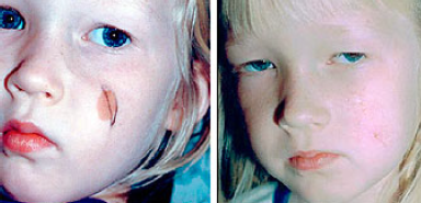
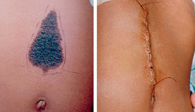
Treatment of congenital nevi depends on size of the lesion, location, estimated risk for melanoma, and expected cosmetic outcome from surgical removal. Although various options for removal exist, surgical removal usually gives the best cosmetic result and the lowest risk for regrowth after treatment.
Giant Congenital Nevi: A Special Scenario

Babies with giant congenital moles clearly have an increased risk of developing melanomas. We estimate that up to 10% of children with giant moles may develop melanoma over a lifetime. Many of these melanomas will occur during the first ten years of life, so that it is important that these giant moles be removed as early as possible. We generally recommend consultation with a pediatric plastic surgeon, beginning at three months of life (or earlier if we are concerned that melanoma is already present). There are a variety of surgical procedures that allow these large areas of skin to be removed and various options for closure of the wound after the mole is removed including skin grafting, artificial skin substitutes and tissue expanders.