Magnetic Resonance Imaging (MRI)
What’s an MRI (Magnetic Resonance Imaging)?
An MRI (magnetic resonance imaging) is a safe and painless test that uses magnets and radio waves to make detailed pictures of the body’s organs, muscles, soft tissues, and structures. Unlike a CAT scan, an MRI doesn’t use radiation.
MRIs are done in hospitals and at radiology centers.
What’s Involved in an MRI?
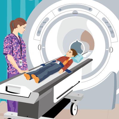
In most MRIs, the scanner consists of a large donut-shaped magnet with a tunnel in the center. This is sometimes called a “closed MRI.” Patients lie on a table that slides into the tunnel. Some centers have MRI scanners with larger openings (an “open MRI”), which are helpful for patients who don’t like tight spaces.
During the test, radio waves and magnets temporarily move the body’s atoms. This is not felt. The movements are picked up by a powerful antenna and sent to a computer. The computer does millions of calculations to create clear, cross-sectional black-and-white images of the body. These images can be converted into three-dimensional (3-D) pictures of the scanned area. These images help to pinpoint problems in the body.
Sometimes, an MRI can provide clear images of body parts that can’t be seen as well with an X-ray, CAT scan, or ultrasound.
Why Are MRIs Done?
Health care providers use MRIs to detect a variety of conditions. The scans can look for problems in the:
- brain and spinal cord
- bones
- joints
- chest
- lungs
- heart and circulatory system
- abdomen and pelvis
- eyes and ears
MRIs highlight contrasts in soft tissue, which helps doctors pinpoint problems with joints, cartilage, ligaments, and tendons. MRI also can help them identify infections and inflammatory conditions, and rule out problems such as tumors.
What Are the Types of MRIs?
Common MRI scans ordered include:

